X-ray (Plain Film)
This machine takes pictures of the inside of your body using a type of radiation called X-rays. These are particularly effective at showing bones but can also show changes in your tissue. You might have an X-ray to look at broken bones, lung conditions, or to check for certain diseases. They are used to quickly help identify an abnormality, e.g. lung conditions or bone fractures.
During the X-ray, you will either stand, sit, or lie down, depending on the area being examined. You will need to keep very still and may be asked to hold your breath for a moment. The process is quick and painless, usually taking just a few seconds.
X-rays involve a small amount of radiation.
You can find out more about general uses of x-rays in radiology here.
Fluoroscopy
Fluoroscopy creates moving images of your body in real time using low-dose X-rays. This can be very helpful for seeing how things work inside you, like your digestive system or blood flow. It’s also used during certain procedures to guide doctors, such as placing catheters.
For the procedure, you may need to stand, sit, or lie on a mobile table. You may be asked to drink a liquid contrast dye beforehand, or this may be given to you via an injection. This is to better define the internal structures being investigated and help diagnose any abnormality.
Fluoroscopy uses X-rays and involves some radiation.
Mammography
A mammogram is a specialist X-ray used to examine breast tissue. It’s a key tool for detecting breast cancer as early as possible and for monitoring other breast conditions. Mammograms can show changes in the breast that are too small to be felt.
During the mammogram, you will stand in front of the machine and your breast will be placed on a flat surface. A plastic plate will press down on your breast to spread the tissue so that a clear image can be scanned. This might feel uncomfortable, but it will only last a few seconds.
Mammograms use a low dose of radiation.
You can learn more about mammograms on the NHS website, and you can read about why the NHS offer breast screenings here.
Computed Tomography (CT)
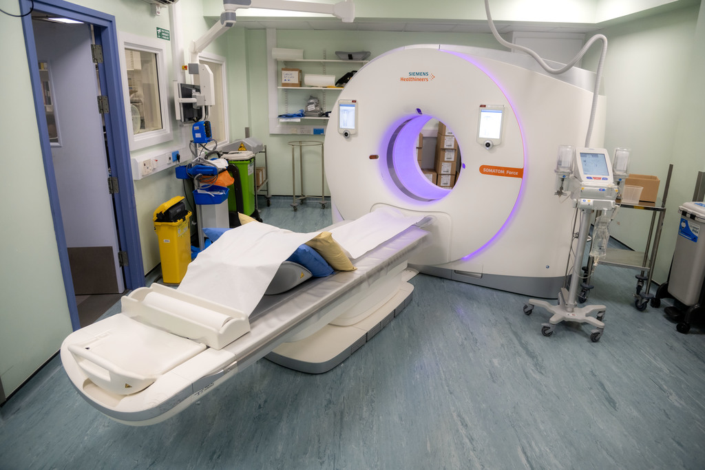
A CT scanner is often referred to as a doughnut. It uses X-rays to take detailed images of many different parts of the body. It can be used to look at bones and soft tissues and is used to diagnose or monitor a range of different illnesses. These include; cardiology; colorectal; gynaecology; musculoskeletal; neurology; urology; and vascular.
During the scan you will need to lie on the scanner bed, keep still and may be asked to hold your breath. The CT scanner is not loud but sounds more like a washing machine spinning. The scans generally only take a few minutes.
For some scans you will be given an injection of contrast; this is a dye that highlights different body tissues to make certain diagnoses easier, but this may cause some side effects, including weakness, sweating and difficulty breathing. In other occasions you may be asked to drink water or oral contrast liquid prior to the scan to help our radiologists. The radiology team will help explain on the day of your appointment.
CT scans use X-rays, which involve a small amount of radiation.
You can find more information on CT scans here.
DEXA (Bone Density Scan)
DEXA stands for Dual-Energy X-ray Absorptiometry. It’s a specialised X-ray study that measures bone mineral density and is important for diagnosing or monitoring conditions like osteoporosis.
You will lie on an open table and the scanner will pass over your body, usually taking about 10-20 minutes.
DEXA scans use very low levels of X-rays, lower than a standard X-ray, making it a safe and valuable tool for assessing bone health.
You can learn more about DEXA scans here.
Ultrasound
Ultrasound uses high-frequency sound waves to create images of the inside of your body. It’s commonly used to look at organs, blood flow, and during pregnancy. It’s also useful for examining soft tissues and for guiding certain procedures.
For the scan, you’ll lie on a bed and a gel will be applied to your skin. The sonographer or radiologist will move a small device called a transducer over the gelled area. The process is painless and usually takes 10-20 minutes.
Ultrasound is very safe and doesn’t use any radiation; the sound waves are harmless and provide a clear picture of what’s going on inside your body.
You can find out more about ultrasounds here.
MRI (Magnetic Resonance Imaging)
MRI uses strong magnets and radio waves to create detailed images of the inside of your body. It’s especially good for looking at soft tissues like the brain, muscles, and organs. MRI is used to diagnose and monitor many conditions, from joint problems to brain disorders.
During the scan, you will lie on a table that slides into a large tube. It’s important to stay very still and sometimes hold your breath for short periods. The MRI machine makes loud knocking noises, but you’ll be given earplugs or headphones to make it the experience more comfortable.
MRI doesn’t use radiation, but the powerful magnets mean you cannot have metal objects with you. It may not be safe to have an MRI if you have certain metals or implantable devices within your body, and our team will check this with you prior to the scan. The scan usually takes 15-45 minutes, depending on what part of your body is being examined.
For more information about MRI scans, please visit this website.
Nuclear Medicine
Nuclear Medicine uses small amounts of radioactive materials to diagnose a variety of diseases. This imaging technique provides detailed pictures of what’s happening inside your body at a cellular level, and is especially effective at detecting conditions like cancer, heart disease, and certain brain disorders.
Before your scan, you will receive a small amount of a radioactive substance, called a tracer, usually through an injection, but sometimes by swallowing or inhaling it. The tracer travels through your body and accumulates in the area being studied. The scan might happen right away or a few hours after the tracer is given, depending on the type of study.
During the scan, you will lie still on a table while a special camera detects the radiation emitted by the tracer and creates images of your internal organs and tissues. The typically takes between 30 minutes to 1 hour.
In other forms of nuclear studies, small amounts of radioactive materials may be combined with CT or MRI scans. This allows better definition of the anatomical areas the tracers accumulate and precisely highlight abnormal activity. These combined investigations (for example in a PET-CT) may be used during cancer investigates to define sites of cancer spread or to confirm successful treatment.
The radiation dose you receive is the same as other imaging tests, such as an X-Ray.
You can find out more about Nuclear Medicine and its use within PET scans here.
Interventional Radiology (IR)
Interventional Radiology (IR) is a specialty that uses imaging guidance, such as X-rays, CT scans or ultrasounds, to perform minimally invasive procedures. These procedures are often used to diagnose or treat a wide range of conditions without the need for traditional surgery. IR can be used to treat blocked blood vessels, insert stents, drain fluid collections, and take tissue samples (biopsies) from organs such as the lymph node or liver.
During an IR procedure, you might be given local anaesthesia to numb the area and sometimes sedation to help you relax. A small incision or needle puncture is made, and the interventional radiologist uses imaging to guide millimetre-wide instruments, such as a catheter or wire, into the area that requires treatment.
The procedures are usually quick and involve less pain and recovery time compared to invasive surgery. You might feel some pressure or discomfort during the procedure, but this is typically not too painful.
IR procedures often use imaging techniques that involve some radiation, but they are carefully planned to ensure the smallest necessary dose is used.
Use of radiation
Some of these scans and procedures involve a small amount of radiation depending on what is being investigated, similar to a of couple days of background radiation or what you would get from commercial flight.
The benefit of getting a clear picture to help diagnose your condition is considered to outweigh any risks, but please speak to one of our radiologists if you have any concerns about this.